
TriTom
Multimodal molecular imaging system for small animal preclinical research
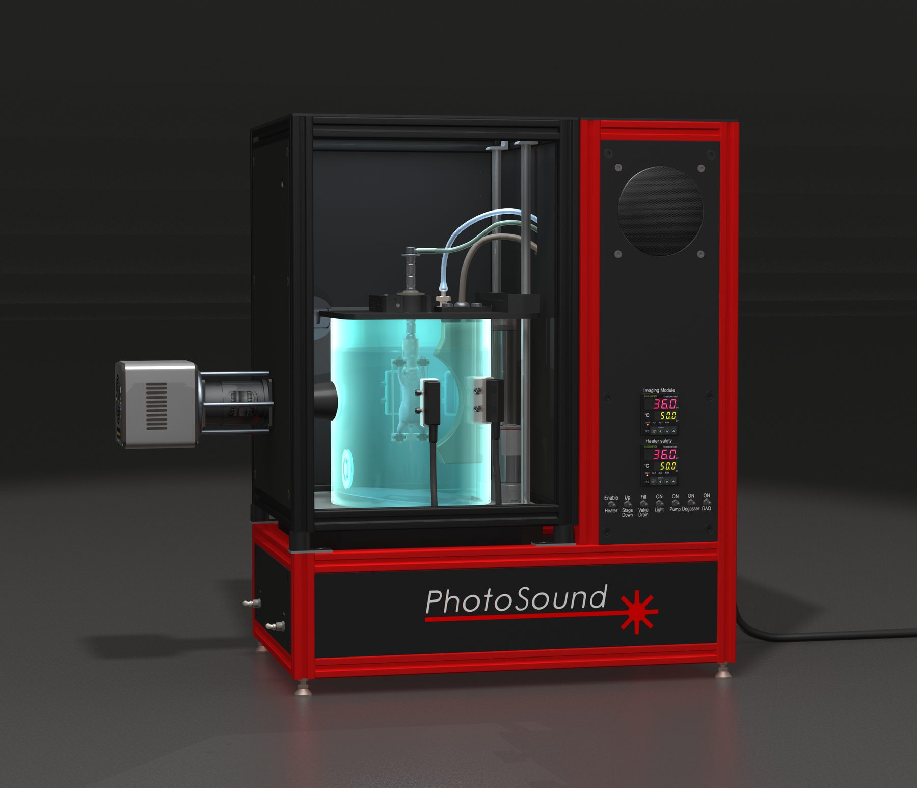
Two modalities, one co-registered volume
TriTom delivers fast, high-resolution 3D imaging by combining photoacoustic and fluorescence tomography in one system. It captures deep-tissue anatomical, functional and molecular data — without the need for contrast agents.
Designed for preclinical researchers, TriTom streamlines in vivo imaging with speed, clarity and precision, so every scan returns a quantitative 3D map registered to the animal's own anatomy.
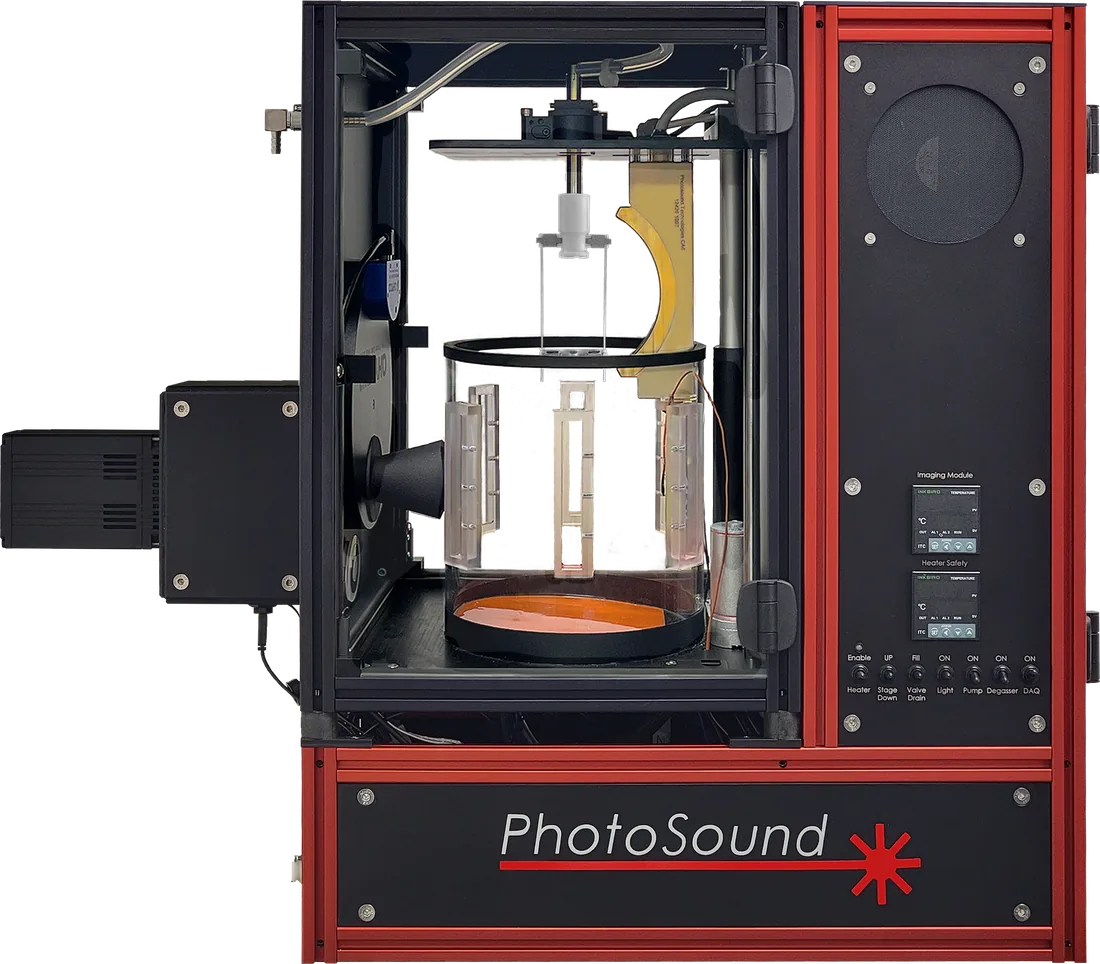
At a glance
- 36 seconds
- A full 360° scan of a 75 cm³ volume; 10 seconds for a local region of interest.
- 150 µm
- Spatial resolution in transverse planes, independent of image size and depth in the object.
- 20–40 mm
- Imaging depth, with no degradation of spatial resolution.
- 460–1320 nm
- Tunable excitation covering visible, NIR I and NIR II probes, switchable every 50 ms.

From power-on to scan in minutes
TriTom is easy to set up, with built-in animal handling, temperature control and intuitive software. No complex calibration or assembly is required.
Holders mount magnetically, anesthesia gas flows continuously through the hollow shaft, and a camera feed lets you check position and animal health while the scan runs. Reconstruction takes under a minute and writes standard volumetric formats you can open in 3D Slicer.
Where researchers use it
Fourteen research areas, from anatomical and cancer imaging through contrast agent development to therapeutic monitoring. Each one links to the matching TriTom publications.